Neurotensin Antibody
ImmunoStar
SKU / CAT#: 20072
$27500
usd$2.75/µL
Neurotensin Antibody
Immunostar Inc.SKU / CAT#: 20072
MPN: 20072
$27500
usd$2.75/µL
Available
SHIPS Jul 17th
Available
SHIPS Jul 17th
SKU
Unit size / Options
Availability
Price
No products found
Description
The rabbit antibody for Neurotensin is generated against synthetic Neurotensin conjugated to bovine thyroglobulin with glutaraldehyde. The antibody is provided as 100 µL of lyophilized whole serum, 0.09% sodium azide.
The rabbit antibody for Neurotensin is generated against synthetic Neurotensin conjugated to bovine thyroglobulin with glutaraldehyde. The antibody is provided as 100 µL of lyophilized whole serum, 0.09% sodium azide.
Shipping & Handling
Shipped In
Ambient
Safety & Storage
Storage Temperature
-20°C
Safety Statement
This product contains the preservative sodium azide. The concentration percent of the sodium azide is ≤ .09%. Although this hazardous substance is a concentration below that required for the preparation of a Material Safety Data Sheet, we created a standard MSDS for your records. This product is intended only for laboratory research and development purposes.
Regulatory & Compliance
Specifications
CLASS
Primary
COUNTRY OF ORIGIN
United States of America
*USAGE / SAFETY STATEMENT
This product contains the preservative sodium azide. The concentration percent of the sodium azide is ≤ .09%. Although this hazardous substance is a concentration below that required for the preparation of a Material Safety Data Sheet, we created a standard MSDS for your records. This product is intended only for laboratory research and development purposes.
REGULATORY NOTICE & RESTRICTIONS
For Laboratory Reagent Use Only. Analytical and performance characteristics are not established. THIS PRODUCT IS FOR RESEARCH USE ONLY AND IS NOT INTENDED FOR DIAGNOSTIC OR THERAPEUTIC USE.
Questions & Answers (0)
Reviews & Ratings (3)
4.7 out of 5 stars
5
67%
4
33%
3
0%
2
0%
1
0%
Staining for Ventromedial Ventral Pallidum
Christina R.
University of California - Irvine
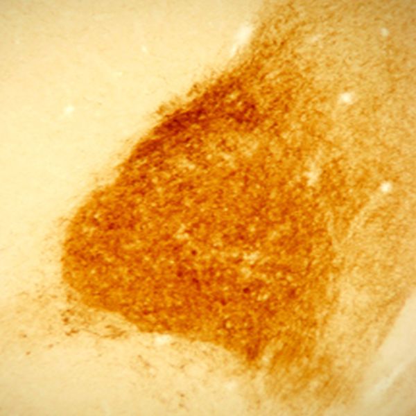
We used the rabbit anti-neurotensin antibody for staining of ventromedial ventral pallidum in rats who were observed during cocaine self administration. Rats were perfused with saline followed by 4% PFA and stored in 20% sucrose-azide. Brains were coronally sectioned to 40 microns and stored in 0.1 M PBS-Azide (pH 7.4). The following protocol for IHC staining was used: Free floating sections of rat brain were blocked in 1% hydrogen peroxide in 0.1M PBS for 15 minutes. After washing, tissue was blocked in 0.1M PBS-Triton X-100 (PBST) and 2% normal donkey serum (NDS) for 2 hours. After washing, primary antibody was used in a range of concentrations (1:2500, 1:5000, 1:10000, 1:20000) overnight in PBST and 2% NDS. After washing, secondary antibody used was biotinylated donkey anti-rabbit (Jackson ImmunoResearch) in a 1:500 dilution in PBST over 2 hours. After wash, tissue placed in ABC peroxide (kit from Vecta Stain) 1:500 in PBST for 1.5 hours. Tissue was developed in brown DAB for 10 minutes. The borders of the ventromedial VP were very clear with this antibody and with low background. Even with concentrations ranging from 1:2500 to 1:20,000, the subregion borders were very easy to identify without having to perform the DAB reaction for an extended period of time.
Visualization of ventromedial ventral pallidum
David R.
Rutgers University
For our manuscript, Differential roles of ventral pallidum subregions during cocaine self-administration behaviors, in press at Journal of Comparative Neurology, we used the Immunostar rabbit anti-neurotensin primary antibody to visualize the ventromedial ventral pallidum subregion. Rats were perfused with saline followed by 4% PF, stored in 30% sucrose, and coronally sectioned to 40 um. We followed the protocols of Zahm/Heimer and colleagues when they discovered the ventral pallidum subregions and their afferent/efferent projection patterns.
The protocol consisted of the following steps:
1. Wash in 0.1M phosphate buffer (PB; pH 7.4)
2. 15 min in 1% sodium borohydride
3. Wash
4. 1 hour blocking with PB containing 0.1% Triton X-100 and 3% normal goat serum
5. Primary antibody overnight at 4C - ImmunoStar rabbit anti-neurotensin diluted 1 : 6500 in PB containing 0.1% Triton X-100 and 3% normal goal serum
6. Wash
7. 1 hour anti-rabbit biotinylated secondary (Vector) 1:200 in PB with 0.1% Triton X-100
8. Wash
9. 1 hour ABC (Vector) 1:200 in PB with 0.1% Triton X-100
10. Wash
11. Develop with 0.05% DAB for 6 min
Our study involved recording neurons within the ventral pallidum subregions during specific aspects of intravenous cocaine self-administration (approaching toward, responding on, or retreating away from a cocaine-reinforced operandum). In order to verify the placement of microwires within the VP subregions, prior to perfusion we passed current through each stainless steel microwire to leave an iron deposit at the uninsulated tip. After DAB (brown reaction) and mounting, we visualized the iron deposit by incubating in a 5% potassium ferrocyanide and 10% HCl solution (leaving a blue-green reaction). For our purposes, this antibody labeled fibers in ventromedial VP with low background. Individual neurons and other fibers were clearly observed within BNST that were not involved in our study. I rated this antibody 4 stars because there was some variability for this stain between animals.
Crisp, Cell-specific labeling
Daniel T.
University of Rochester
Tissue: Maccaca fasciscularis, perfused with saline and 4% paraformaldehyde, dehydrated through increase sucrose gradients, sectioned at 40 um with sliding microtome. Method: Immunocytochemistry on free floating sections. Incubated with ImmunoStar's primary antibody at a concentration of 1:1000 for 4 days in 10% normal goat serum (in 0.1M PO4 buffer with .3% Triton X-100). Rinsed, then incubated with Vector Lab's biotinylated goat anti-rabbit secondary antibody (#BA-1000) at a concentration of 1:200 for 40 minutes at room temperature in the same 10% normal goat serum solution. Rinsed, then incubated with Vector's standard peroxidase kit (#PK-4000) for 60 minutes at room temperature in 0.1M PO4 buffer with .3% Triton X-100. Rinsed, then developed using the instructions for the kit. Results: Crisp, cell-specific labeling. Low background. Very helpful for determining the boundary between the medial and lateral core divisions of the central nucleus of the amygdala.
Citations (0)

Shipping & Handling
Shipped In
Ambient
Safety & Storage
Storage Temperature
-20°C
Safety Statement
This product contains the preservative sodium azide. The concentration percent of the sodium azide is ≤ .09%. Although this hazardous substance is a concentration below that required for the preparation of a Material Safety Data Sheet, we created a standard MSDS for your records. This product is intended only for laboratory research and development purposes.
Regulatory & Compliance
Join Our List

Subscribe to the Scoop blog and get the latest updates on life in the lab.
Related Blog Articles
Related Blog Articles