Specifications
CLASS
Optical Clearing Agent
*USAGE / SAFETY STATEMENT
For Research and Laboratory Use Only.
APPLICATION
Animal: brain, kidney, heart, stomach, intestine, liver, lung, pancreas, skin; Insect: brain, ventral nerve cord, ovary; Biomaterial: collagen matrix; Plant
CLEARING SOLITARY USE
Recommended 0.5~1mm depth tissue slice
CLEARING WITH CLARITY
Recommended Whole Organ
CLEARING WITH SCALEVIEW
Not Recommended
CLEARING SPEED SOLITARY USE
Within 1 Hour
CLEARING SPEED WITH CLARITY
Days
IMMUNOSTAINING COMPATIBILITY
Yes
FLUORESCENT PROTEIN COMPATIBILITY
Yes
ORGANIC DYE COMPATIBILITY
Yes
LIPOPHILIC DYE COMPATIBILITY
Yes
OBJECTIVE LENS
Objective lens with high NA and long WD is suggested:
Leica HCX IRAPOL 25X water-immersion objective, NA 0.95, WD 2.4mm;
Zeiss 20X W-PlanApochromat objective, NA 1.0, WD 2.0 mm
Olympus XLSLPLN25XSVMP objective, NA 0.9, WD 8.0 mm
PROPERTIES
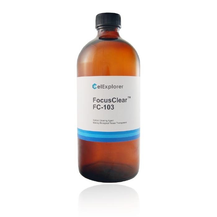
FocusClearTM solution is a water-soluble clearing agent. It is not gelling in the bottle and no dehydration of the objects is necessary. Samples can be directly transferred from water, buffer solutions, alcohol, DMSO, DMF, and glycerin into FocusClearTM solution. FocusClearTM can be used for samples labeled with fluorescence and non-fluorescence dyes including lipophilic dyes, such as DiI, DiD and NBD-ceramide. FocusClearTM is non-toxic, ready to use, always liquid, no need to be aliquoted, mixed, centrifuged or kept frozen. It allows easy and universal production of preparations. MountClear TM is a mountant which specially designed for mounting specimens cleared by the FocusClearTM. MountClear TM does not interfere the clearing effect of FocusClear TM. In addition, it has anti-quenching, non-fluorescence and quick clotting characteristics. Using mounting media other than MountClearTM may result in cloudiness of the sample. Immersion Solution-M is an immersion solution with a refraction index matching to those of MountClear TM. They are designed to avoid deformation of the observed images during high-resolution microscopic observation that using glycerin or water immersion objective lens. Effects: Tissues in the FocusClearTM become transparent. The resolution and depth of focus greatly increased with sharp outline and high contrast. In order to obtain best results, it is recommended that the specimen cleared in FocusClearTM should be mounted in MountClearTMand observed with oil or water immersion lens with high numerical aperture and covered with Immersion Solution-M. FocusClearTM, however, is designed to clear specimens fixed by cross-linking agents such as paraformaldehyde and glutaraldehyde, but it is ineffective for heat-denatured or alcohol fixed specimens.
EFFECTS
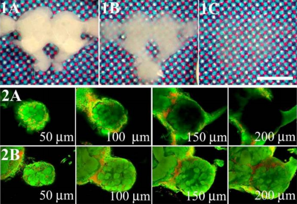
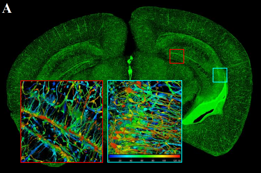
Tissues in the FocusClearTM become transparent. The resolution and depth of focus greatly increased with sharp outline and high contrast. In order to obtain best results, it is recommended that the specimen cleared in FocusClearTM should be mounted in MountClearTMand observed with oil or water immersion lens with high numerical aperture and covered with Immersion Solution-M. FocusClearTM, however, is designed to clear specimens fixed by cross-linking agents such as paraformaldehyde and glutaraldehyde, but it is ineffective for heat-denatured or alcohol fixed specimens.
APPLICATION PROTOCOL
1. Paraformaldehyde and/or glutaraldehyde fixed samples labeled with immunofluorescence, fluorescence probes, immunohistochemicals, or conventional dyes should be thoroughly washed to remove non-specific bindings.
2. Tissue blocks, brain slices, cryosections or single cells ready for microscopic observation can be directly transferred into appropriate amount of FocusClearTM solution for clearing. Note: For an intact fly brain, 100µl FocusClearTM solution is suggested. For a slice of mouse brain (200 µm thick), 1 ml FocusClearTM solution is suggested.
3. For an effective clearing, the incubation time (10 min to 4 h) should be adjusted according to the size of the tissue (106µm3 ~ 1 mm3). To prevent evaporation during clearing, the incubation chamber should be completely sealed with parafilm membrane. Note: Small samples such as fly brains may become completely transparent and difficult to be retrieved under a dissecting microscope. By simply applying a drop of saline solution, your precious samples will become visible again. You can clear the tissue again in a smaller drop of FocusClearTM for easy retrieval.
4. The cleared specimens are then mounted in a fresh drop of FocusClearTM solution.
5. For the best quality, the cleared specimens should be mounted in the MountClearTM solution. Prior to every use, the MountClearTM solution should be completely dissolved again by incubation in the hot-water bath (55℃) for about 30 min. After brief cooling at room temperature, the MountClearTM solution is ready for use.
6. Seal the sample completely with fingernail polisher.
7. When using an oil/water immersion lens to observe the sample, Immersion Solution-M matching the reflective index of the mounting solution should be used for better resolution.


